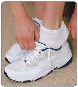
How Shoe Lifts Overcome Leg Length Difference
There are not one but two unique variations of leg length discrepancies, congenital and acquired. Congenital means that you are born with it. One leg is anatomically shorter compared to the other. As a result of developmental phases of aging, the brain senses the step pattern and recognizes some variation. The human body typically adapts by tilting one shoulder over to the "short" side. A difference of less than a quarter inch is not very excessive, demand Shoe Lifts to compensate and commonly won't have a profound effect over a lifetime.

Leg length inequality goes largely undiscovered on a daily basis, yet this issue is very easily fixed, and can eradicate many instances of upper back pain.
Therapy for leg length inequality usually consists of Shoe Lifts. Most are low-priced, usually costing less than twenty dollars, in comparison to a custom orthotic of $200 or more. Differences over a quarter inch can take their toll on the spine and should probably be compensated for with a heel lift. In some cases, the shortage can be so extreme that it requires a full lift to both the heel and sole of the shoe.
Low back pain is the most common ailment afflicting people today. Around 80 million people are afflicted by back pain at some stage in their life. It is a problem that costs companies huge amounts of money yearly because of time lost and output. New and superior treatment solutions are constantly sought after in the hope of lowering economic impact this issue causes.

Men and women from all corners of the earth suffer the pain of foot ache as a result of leg length discrepancy. In these types of situations Shoe Lifts can be of worthwhile. The lifts are capable of eliminating any pain and discomfort in the feet. Shoe Lifts are recommended by numerous professional orthopaedic physicians.
In order to support the body in a nicely balanced fashion, your feet have a significant part to play. Despite that, it is sometimes the most neglected region in the body. Some people have flat-feet which means there is unequal force exerted on the feet. This will cause other areas of the body including knees, ankles and backs to be affected too. Shoe Lifts ensure that proper posture and balance are restored.
Leg length inequality goes largely undiscovered on a daily basis, yet this issue is very easily fixed, and can eradicate many instances of upper back pain.
Therapy for leg length inequality usually consists of Shoe Lifts. Most are low-priced, usually costing less than twenty dollars, in comparison to a custom orthotic of $200 or more. Differences over a quarter inch can take their toll on the spine and should probably be compensated for with a heel lift. In some cases, the shortage can be so extreme that it requires a full lift to both the heel and sole of the shoe.
Low back pain is the most common ailment afflicting people today. Around 80 million people are afflicted by back pain at some stage in their life. It is a problem that costs companies huge amounts of money yearly because of time lost and output. New and superior treatment solutions are constantly sought after in the hope of lowering economic impact this issue causes.
Men and women from all corners of the earth suffer the pain of foot ache as a result of leg length discrepancy. In these types of situations Shoe Lifts can be of worthwhile. The lifts are capable of eliminating any pain and discomfort in the feet. Shoe Lifts are recommended by numerous professional orthopaedic physicians.
In order to support the body in a nicely balanced fashion, your feet have a significant part to play. Despite that, it is sometimes the most neglected region in the body. Some people have flat-feet which means there is unequal force exerted on the feet. This will cause other areas of the body including knees, ankles and backs to be affected too. Shoe Lifts ensure that proper posture and balance are restored.
The Best Way To Protect Against Calcaneal Spur
Overview
Heel Spurs should be called a traction spurs because they grow in the same direction that the tendons pull away from the heel bone, which is why it can occur on the bottom of the heel (Plantar Fasciitis) and on the back of the heel (Achilles Tendonitis). Some patients may only develop one type of heel spur, but both these problems are closely related so it's not unusual for a patient to have both heel spurs. It's important to note though that most heel spurs aren't the cause of your heel pain.
Causes
Bone spurs form in the feet in response to tight ligaments, to activities such as dancing and running that put stress on the feet, and to pressure from being overweight or from poorly fitting shoes. For example, the long ligament on the bottom of the foot (plantar fascia) can become stressed or tight and pull on the heel, causing the ligament to become inflamed (plantar fasciitis). As the bone tries to mend itself, a bone spur can form on the bottom of the heel (known as a ?heel spur?). Pressure at the back of the heel from frequently wearing shoes that are too tight can cause a bone spur on the back of the heel. This is sometimes called a ?pump bump,? because it is often seen in women who wear high heels.

Symptoms
Heel spurs result in a jabbing or aching sensation on or under the heel bone. The pain is often worst when you first arise in the morning and get to your feet. You may also experience pain when standing up after prolonged periods of sitting, such as work sessions at a desk or car rides. The discomfort may lessen after you spend several minutes walking, only to return later. Heel spurs can cause intermittent or chronic pain.
Diagnosis
A heel spur is often seen on X-ray as a bony protrusion, which can vary in size. However, because a Heel Spur only indicates increased load on the plantar fascia, and not pain, an ultra sound may be required to assess other actual cause of the heel pain such and may include checking to see if the plantar fascia is inflamed or degenerated.
Non Surgical Treatment
Perform some exercises. Exercises that strengthen and lengthen your plantar fascia can also be very helpful for heel spurs. Try some of the following activities. Calf stretch. Place your hands on a wall. Extend 1 foot (0.3 m) behind you with your knee straight and place the other foot in front of you with the knee bent. Push your hips toward the wall and hold the stretch for 10 seconds. You should feel a pull in your calf muscles. Repeat the stretch 20 times for each foot. Plantar fascia stretch, Perform this exercise in the morning before you've done any standing or walking. Cross your injured foot over the knee of your other leg. Grasp your toes and gently pull them toward you. If you can't reach your toes, then wrap a towel around them and pull on the towel. Hold the stretch for 10 seconds and repeat 20 times for each foot.
Surgical Treatment
Surgery is used a very small percentage of the time. It is usually considered after trying non-surgical treatments for at least a year. Plantar fascia release surgery is use to relax the plantar fascia. This surgery is commonly paired with tarsal tunnel release surgery. Surgery is successful for the majority of people.
What May Cause Posterior Calcaneal Spur

Overview
Heel spurs are a condition that usually makes its presence known first thing in the morning via heel pain. Discomfort is typically felt in the front and bottom of the heel (calcaneal). Pain can be constant for several months or intermittent for lengthy periods of time.
Causes
Bone spurs can form anywhere in the feet in response to tight ligaments, repetitive stress injuries (typically from sports), obesity, even poorly fitting shoes. For instance, when the plantar fascia on the bottom of the foot pulls repeatedly on the heel, the ligament becomes inflamed, causing plantar fasciitis. As the bone tries to mend itself, a bone spur forms on the bottom of the heel, typically referred to as a heel spur. This is a common source of heel pain.

Symptoms
Heel spur and plantar fasciitis pain usually begins in the bottom of the heel, and frequently radiates into the arch. At times, however, the pain may be felt only in the arch. The pain is most intense when first standing, after any period of rest. Most people with this problem experience their greatest pain in the morning, with the first few steps after sleeping. After several minutes of walking, the pain usually becomes less intense and may disappear completely, only to return later with prolonged walking or standing. If a nerve is irritated due to the swollen plantar fascia, this pain may radiate into the ankle. In the early stages of Heel Spurs and Plantar Fasciitis, the pain will usually subside quickly with getting off of the foot and resting. As the disease progresses, it may take longer periods of time for the pain to subside.
Diagnosis
A Heel Spur diagnosis is made when an X-ray shows a hook of bone protruding from the bottom of the foot at the point where the plantar fascia is attached to the heel bone. The plantar fascia is the thick, connective tissue that runs from the calcaneus (heel bone) to the ball of the foot. This strong and tight tissue helps maintain the arch of the foot. It is also one of the major transmitters of weight across the foot as you walk or run. In other words, tremendous stress is placed on the plantar fascia.
Non Surgical Treatment
The key for the proper treatment of heel spurs is determining what is causing the excessive stretching of the plantar fascia. When the cause is over-pronation (flat feet), an orthotic with rearfoot posting and longitudinal arch support is an effective device to reduce the over-pronation, and allow the condition to heal. Other common treatments include stretching exercises, losing weight, wearing shoes that have a cushioned heel that absorbs shock, and elevating the heel with the use of a heel cradle, heel cup, or orthotic. Heel cradles and heel cups provide extra comfort and cushion to the heel, and reduce the amount of shock and shear forces experienced from everyday activities.
Surgical Treatment
Have surgery if no other treatments work. Before performing surgery, doctors usually give home treatments and improved footwear about a year to work. When nothing else eases the pain, here's what you need to know about surgical options. Instep plantar fasciotomy. Doctors remove part of the plantar fascia to ease pressure on the nerves in your foot. Endoscopy. This surgery performs the same function as an instep plantar fasciotomy but uses smaller incisions so that you'll heal faster. However, endoscopy has a higher rate of nerve damage, so consider this before you opt for this option. Be prepared to wear a below-the-knee walking cast to ease the pain of surgery and to speed the healing process. These casts, or "boots," usually work better than crutches to speed up your recovery time.
Prevention
A variety of steps can be taken to avoid heel pain and accompanying afflictions. Wear shoes that fit well-front, back, and sides-and have shock-absorbent soles, rigid shanks, and supportive heel counters. Wear the proper shoes for each activity. Do not wear shoes with excessive wear on heels or soles. Prepare properly before exercising. Warm up and do stretching exercises before and after running. Pace yourself when you participate in athletic activities. Don't underestimate your body's need for rest and good nutrition. If obese, lose weight.
Cure For Bursitis Of The Foot
Overview
Infracalcaneal bursitis (inflammation of the bursa below the calcaneus, or heel bone) is one of the most common types of bursitis in the foot. Infracalcaneal bursitis can sometimes be difficult to differentiate from plantar fasciosis-another condition that causes pain below the heel. The key difference is that infracalcaneal bursitis tends to be worse at the end of the day whereas plantar fascia pain tends to be worse in the morning, immediately upon waking.
Causes
A rapid increase in physical activity levels or thinning of the heel?s protective fat pad are factors that may contribute to infracalcaneal bursitis. Other possible causes of infracalcaneal bursitis include blunt force trauma, acute or chronic infection, and arthritic conditions. The following factors may increase your risk of experiencing bursitis, including infracalcaneal bursitis. Participating in contact sports. Having a previous history of bursitis in any joint. Poor conditioning. Exposure to cold weather. Heel striking when running, especially in conventional running shoes with heel elevation.
Symptoms
Achiness or stiffness in the affected joint. Worse pain when you press on or move the joint. A joint that looks red and swollen (especially when the bursae in the knee or elbow are affected). A joint that feels warm to the touch, compared to the unaffected joint, which could be a sign that you have an infection in the bursa. A ?squishy? feeling when you touch the affected part. Symptoms that rapidly reappear after an injury or sharp blow to the affected area.
Diagnosis
A good clinical practise includes evaluation of the tendon, bursa and calcaneum by, careful history, inspection of the region for bony prominence and local swelling as well as palpation of the area of maximal tenderness. Biomechanical abnormalities, joint stiffness and proximal soft tissue tightening can exacerbate an anatomical predisposition to retrocalcaneal bursitis, they warrant correction when present.
Non Surgical Treatment
Rest and apply cold therapy or ice. Ice should not be applied directly to the skin as it may cause ice burns but wrap in a wet tea towel. Commercially available hot and cold packs are often more convenience than using ice. Taping the bursa with a donut shaped pad to take some of the pressure from footwear may help. A doctor may prescribe anti-inflammatory medication e.g. ibuprofen to reduce the pain and inflammation. Applying electrotherapy such as ultrasound may reduce inflammation and swelling. A steroid injection followed by 48 hours rest may be given for persistent cases. If the bursitis is particularly bad and does not respond to conservative treatment then surgery is also an option.
Surgical Treatment
Bursectomy is a surgical procedure used to remove an inflamed or infected bursa, which is a fluid-filled sac that reduces friction between tissues of the body. Because retrocalcaneal bursitis can cause chronic inflammation, pain and discomfort, bursectomy may be used as a treatment for the condition when it is persistent and cannot be relived with other treatments. During this procedure, a surgeon makes small incisions so that a camera may be inserted into the joint. This camera is called an arthroscope. Another small incision is made so that surgical instruments can be inserted to remove the inflamed bursa.
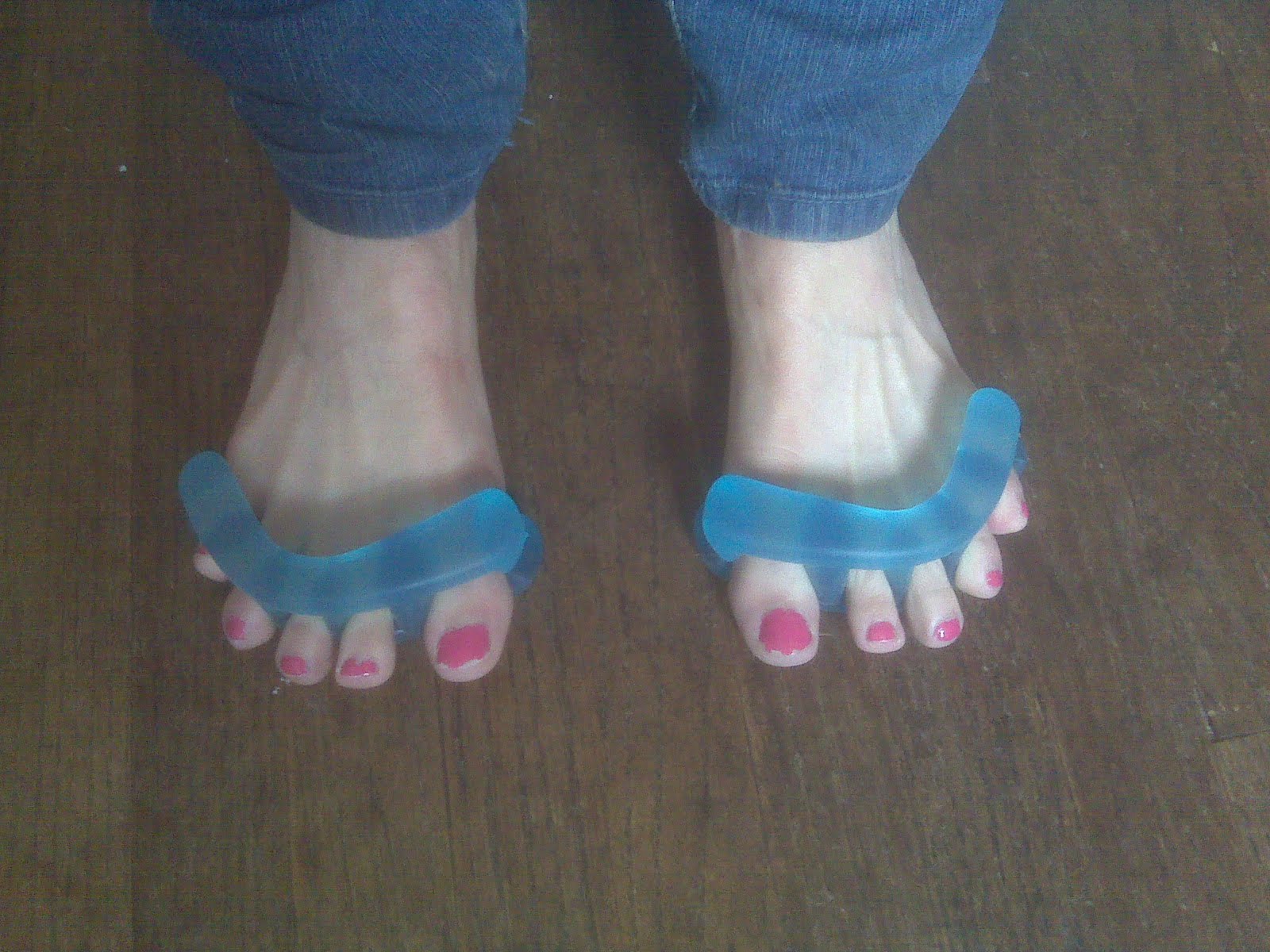
Hammertoes Treatment
 Overview
Hammer toe (hammertoe) is a deformity of the second, third, or fourth toes. In this condition, the toe is bent at the middle joint, into an upward position, causing it to resemble a hammer (sometimes decribed as ?curled toes?). Left untreated, hammer toes can become inflexible and require surgery. Toes which take on a curled appearance are hammer toes. Mallet toe is a similar condition, but affects the upper joint of a toe.
Causes
A hammer toe develops because of an abnormal balance of the muscles in the toes. This abnormal balance causes increased pressures on the tendons and joints of the toe, leading to its contracture. Heredity and trauma can also lead to the formation of a hammer toe. Arthritis is another factor, because the balance around the toe in people with arthritis is disrupted. Wearing shoes that are too tight and cause the toes to squeeze can also cause a hammer toe to form.
 Symptoms
Symptoms of a hammertoe are usually first noticed as a corn on the top of the toe or at the tip which produces pain with walking or wearing tight shoes. Most people feel a corn is due to a skin problem on their toes, which in fact, it is protecting the underlying bone deformity. A corn on the toe is sometimes referred to as a heloma dura or heloma durum, meaning hard corn. This is most common at the level of the affected joint due to continuous friction of the deformity against your shoes.
Diagnosis
Your healthcare provider will examine your foot, checking for redness, swelling, corns, and calluses. Your provider will also measure the flexibility of your toes and test how much feeling you have in your toes. You may have blood tests to check for arthritis, diabetes, and infection.
Non hammertoe Surgical Treatment
Apply a commercial, nonmedicated hammertoe pad around the bony prominence of the hammertoe. This will decrease pressure on the area. Wear a shoe with a deep toe box. If the hammertoe becomes inflamed and painful, apply ice packs several times a day to reduce swelling. Avoid heels more than two inches tall. A loose-fitting pair of shoes can also help protect the foot while reducing pressure on the affected toe, making walking a little easier until a visit to your podiatrist can be arranged. It is important to remember that, while this treatment will make the hammertoe feel better, it does not cure the condition. A trip to the podiatric physician?s office will be necessary to repair the toe to allow for normal foot function. Avoid wearing shoes that are too tight or narrow. Children should have their shoes properly fitted on a regular basis, as their feet can often outgrow their shoes rapidly. See your podiatric physician if pain persists.
Surgical Treatment
Surgery to correct for a hammertoe may be performed as a day procedure. There are several different types of procedures that can be used depending on the foot structure and if the deformity is flexible or rigid.
 Prevention
Custom orthotics paired with a well made shoe can prevent the progression and development of hammertoes. Wearing proper-fitting shoes and custom orthotic devices can provide the support patients need to address muscle/tendon dysfunction. It can also support end stage diseases that result in hammertoe deformities by re-balancing the foot and ankle and controlling the deforming forces.
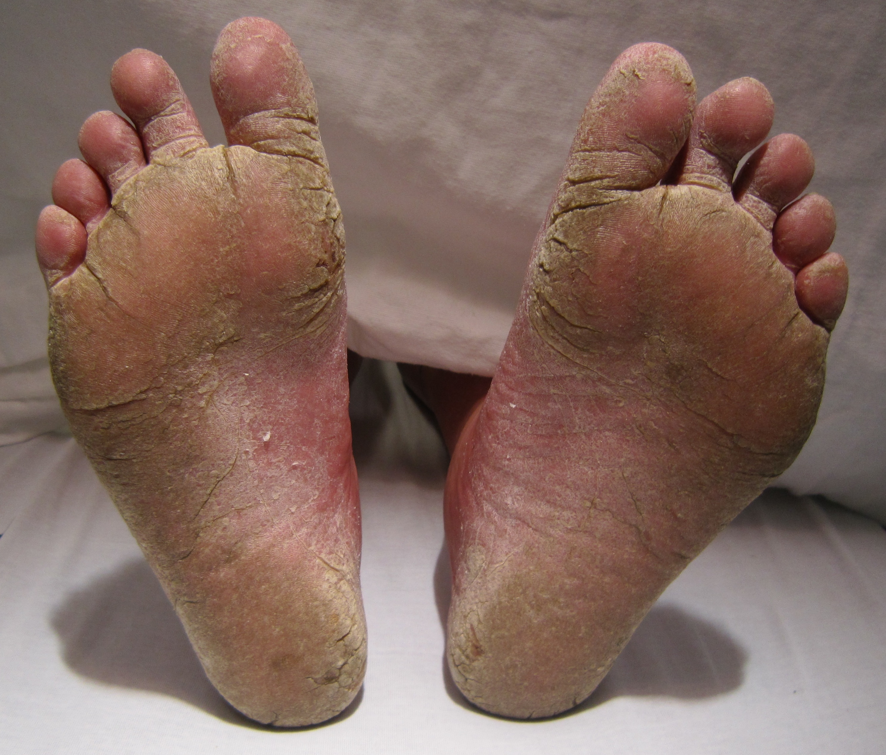
Will Overpronation Of The Feet Involve Surgical Treatments
Overview
Over-pronation, or flat feet, occurs in the walking process when a person?s arch collapses upon weight bearing. This motion can cause extreme stress or inflammation on the plantar fascia, possibly causing severe discomfort and leading to other foot problems. Bear in mind that people with flat feet often do not experience discomfort immediately, and some never suffer from any discomfort at all. Over-pronation can often lead to conditions such as plantar fasciitis, heel spurs, metatarsalgia, post-tib tendonitis, bunions.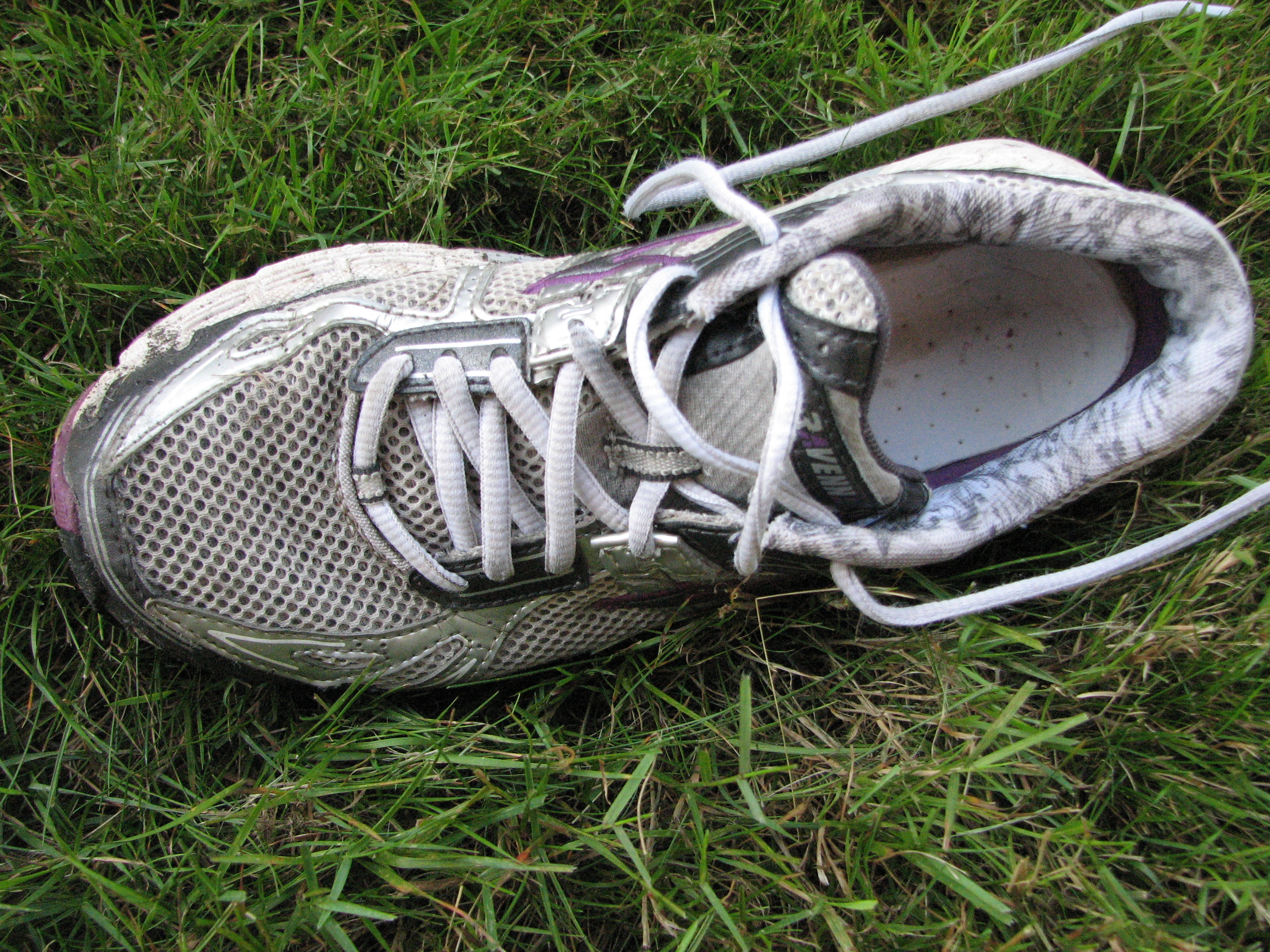
Causes
Excess stress on the inner surface of the foot can cause injury and pain in the foot and ankle. Repeated rotational forces through the shin, knee, thigh and pelvis also place additional strain on the muscles, tendons and ligaments of the lower leg.
Symptoms
Symptoms can manifest in many different ways. The associated conditions depend on the individual lifestyle of each patient. Here is a list of some of the conditions associated with over Pronation. Hallux Abducto Valgus (bunions). Hallux Rigidus (stiff 1st toe). Arch Pain. Heel Pain (plantar Facsitus). Metatarsalgia (ball of the foot pain). Ankle Sprains. Shin Splints. Achilles Tendonitis. Osteochondrosis. Knee Pain. Corns & Calluses. Flat Feet. Hammer Toes.
Diagnosis
When you overpronate your foot rolls inwards causing the lower leg to rotate inwards too. It's thought that this increases stress on the soft tissues of the lower leg causing pain and inflammation, or more commonly known as shin splints.
Non Surgical Treatment
Wear shoes with straight or semicurved lasts. Motion-control or stability shoes with firm, multidensity midsoles and external control features that limit pronation are best. Over-the-counter orthotics or arch supports can help, too. You know you are making improvements when the wear pattern on your shoes becomes more normal. Overpronation causes extra stress and tightness to the muscles, so do a little extra stretching.
Prevention
Custom-made orthotics supports not only the arch as a whole, but also each individual bone and joint that forms the arch. It is not enough to use an over-the-counter arch support, as these generic devices will not provide the proper support to each specific structure of the arch and foot. Each pronated foot?s arch collapses differently and to different degrees. The only way to provide the support that you may need is with a custom-made device. This action of the custom-made orthotic will help to prevent heel spurs, plantar fasciitis, calluses, arch pain, and weakness of the entire foot.
Living With Severs Disease
Overview
Sever?s disease is particularly prevalent among active children between ages 8 and 15. Young boys and girls who play soccer and other sports in which footwear is inappropriate-i.e. too narrow in the toe box, too rigid, etc. are most commonly affected. Sever?s disease usually appears during the adolescent growth spurt-the 2-year period in early puberty where children grow the quickest. The adolescent growth spurt occurs between the ages of 8 and 13 in girls and 10 and 15 in boys. Teenagers over 15 years old rarely experience this heel problem, as heel bone growth is usually complete by this age. Sever?s disease usually self-resolves within 6 months of onset, though it can last longer.
Causes
There are many biomechanical factors that predispose a young athlete to calcaneal apophysitis. The majority of patients will present with an ankle equinus deformity, which ultimately exerts an increased pulling force to the Achilles insertion and non-ossified apophysis. Furthermore, patients may present with hyperpronation of the rearfoot. This allows more of a ?teeter-totter? effect or lack of motion control on the frontal plane of the calcaneus.
Symptoms
Symptoms include Heel Pain. Pain at the back of the heels when walking or running. Possibly a lump at the back of the heel, although this might be minimal. Pain and tenderness at the back of the heels, especially if you press on it, or give it a squeeze from the sides. Tight calf muscles resulting in reduced ankle range of motion.
Diagnosis
Sever disease is most often diagnosed clinically, and radiographic evaluation is believed to be unnecessary by many physicians, but if a diagnosis of calcaneal apophysitis is made without obtaining radiographs, a lesion requiring more aggressive treatment could be missed. Foot radiographs are usually normal and the radiologic identification of calcaneal apophysitis without the absence of clinical information was not reliable.
Non Surgical Treatment
Massage the calves gently from the knee to the heel, being especially careful around the Achilles? tendon, as this will be extremely tight and tender. During this massage, flex and point the foot through normal pain-free ranges of motion to increase flexibility while massaging. Massage every other or every third day, making sure your young athlete is not still sore before massaging again. If you?re unsure how to massage, find someone in your area that uses Graston technique or Active Release Therapy for best results. Stretch your athlete?s calves. This is the most overlooked aspect of treatment for Sever?s Disease and this needs to be done every day after practice, and when first starting we recommend 2-3 times per day, allowing gravity to pull heel down, never forcing the stretch. Ice your heels, but don?t just put an ice pack there. Use a cold water soak to fully immerse the foot and calves up to the knee. We recommend using a rubbermaid can found here. Soak for 10-15 minutes. The water does not have to be frigid, just cold. Use cold water from the tap, insert the foot, then add some ice to help bring down the temperature. When your athlete is experiencing pain, ice every hour, on the hour, for as many times as possible in one day. Make sure the heel/calves are body temperature before beginning again. Support the arches. This is what has been shown in studies to reduce pain in young athletes with Sever?s Disease. If you miss out on this one, you miss out on relieving your athletes pain.
Recovery
Recovery time will vary from patient to patient. Age, health, previous injuries, and severity of symptoms will affect recovery time. Your compliance with the stretching program and the other recommendations made by your doctor will also determine your healing time. Heel pain often completely resolves after a child?s heel bone has stopped its period of growth.